In this blog for men making decisions about having a prostate biopsy, Fransisco Lopez and Alastair Lamb talk about benefits, harms and targeting the cancer that matters.
Page last checked 21 April 2023
Take-home points
- There are prostatic tumours that don’t represent any threat to men, while others are aggressive and could harm.
- Magnetic resonance imaging can help to identify tumours, guiding biopsies to target high riskA way of expressing the chance of an event taking place, expressed as the number of events divided by the total number of observations or people. It can be stated as ‘the chance of falling were one in four’ (1/4 = 25%). This measure is good no matter the incidence of events i.e. common or infrequent. tumours, while avoiding biopsies in those of no clinical significance.
- In patients with a negative MRI and a low suspicion of prostate cancer, prostate biopsy could be avoided based on shared decision-making with their physicians.
Prostate cancer is a disease with multiple behaviours, ranging from small tumours which are clinically meaningless and don’t represent any threat to men, to aggressive lesions that are prone to spread to different parts of the body and, ultimately, cause death. Each prostate cancer has characteristics of its own that can be identified at diagnosis in order to predict behaviour, and certain characteristics found on the tissue obtained on prostate biopsies are among the most significant.
Today, one of the most important challenges in prostate cancer diagnosis is the need to identify only those tumours of clinical significance while avoiding picking up those that are not significant. The detection and treatmentSomething done with the aim of improving health or relieving suffering. For example, medicines, surgery, psychological and physical therapies, diet and exercise changes. of low risk cancers could actually be considered to be a burden, not a benefit, and adds to both healthcare costs and complication rates.
Looking for a needle in a haystack
Undertaking a prostate biopsy might be compared to looking for a needle in a haystack, as multiple systematic samples of the prostate are taken trying to hit a tumour that may be only couple millimetres in size. Interestingly, the haystack can have many needles in it, because prostate cancer can have multiple tumours of varying severity present at the same time. However, usually one of these tumours is more aggressive than the others and defines the course of the disease. This lesion is the one we want to sample.
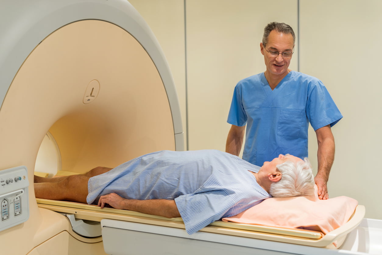
Historically, no diagnostic imaging has been proved useful to identify significant tumours when doing prostate biopsies. Therefore, these have been done without any guidance in a systematic fashion, except when there was an abnormality on rectal examination and could be used to help find a larger tumour. However, recent advances in magnetic resonance imaging (MRI) techniques allow non-invasive identification of prostatic tumours. This has created the possibility of targeting biopsies to suspicious lesions in the prostate rather than taking blind samples of it.

Risks related to prostate biopsy
As with any medical interventionA treatment, procedure or programme of health care that has the potential to change the course of events of a healthcare condition. Examples include a drug, surgery, exercise or counselling. , this procedure has risks that have to be weighed against the benefits. Complications related to prostate biopsy include urinary and rectal bleeding, urinary symptoms, urinary retention (inability to void), transient erectile dysfunction, pain and infections that occasionally might be severe. Therefore, it is very important to identify which patients really need the biopsy and when it can be safely avoided. Overuse of biopsy might result in more harm than good, for example overdiagnosis of cancers that are not agressive and consequent overtreatment due to anxiety or poor counselling. Prostate biopsy is therefore best performed only when there is suspicion of prostate cancer that might cause harm to the patient and MRI is critical in making this decision.
Role of magnetic resonance imaging (MRI) on the diagnosis of prostate cancer
The most straightforward role of MRI in prostate cancer is tumour identification, however, it has much more to offer than just showing us the location of lesions. Some studies have indicated that MRI can be a useful tool to recognise the biological behaviour of prostatic tumours, highlighting those that can be aggressive (Beksac et al, 2018). The combination of these capabilities can help us to reduce the number of biopsy procedures, guiding biopsies to target high risk tumours, while avoiding those of no clinical significance.
A recent Cochrane ReviewCochrane Reviews are systematic reviews. In systematic reviews we search for and summarize studies that answer a specific research question (e.g. is paracetamol effective and safe for treating back pain?). The studies are identified, assessed, and summarized by using a systematic and predefined approach. They inform recommendations for healthcare and research. that included 43 studies found that the introduction of MRI as part of the prostate cancer diagnostic pathway is better than current practice, and may lead to a 12% improvement in diagnostic accuracy (Drost et al., 2019). Although these numbers represent a significant improvement, the authors rated the quality of evidenceThe certainty (or quality) of evidence is the extent to which we can be confident that what the research tells us about a particular treatment effect is likely to be accurate. Concerns about factors such as bias can reduce the certainty of the evidence. Evidence may be of high certainty; moderate certainty; low certainty or very-low certainty. Cochrane has adopted the GRADE approach (Grading of Recommendations Assessment, Development and Evaluation) for assessing certainty (or quality) of evidence. Find out more here: https://training.cochrane.org/grade-approach for these findings as low and that could change with higher quality research .
One of the largest clinical trialsClinical trials are research studies involving people who use healthcare services. They often compare a new or different treatment with the best treatment currently available. This is to test whether the new or different treatment is safe, effective and any better than what is currently used. No matter how promising a new treatment may appear during tests in a laboratory, it must go through clinical trials before its benefits and risks can really be known. addressing the use of MRI in prostate cancer diagnosis found that biopsy might be spared in 27% of patients and that 5% fewer non-clinically significant cancers would be diagnosed (Ahmed et al, 2017). Also, this approach seems to be safe. In a pooled analysis of several publications performed by our group, we found that only 11% of lesions classified as non suspicious on the MRI prove to be of clinical importance if biopsy is still performed (Sathianathen, 2019).
If my MRI was negative, should I get a biopsy?
The probability of diagnosing a clinically significantClinical significance is the practical importance of an effect (e.g. a reduction in symptoms); whether it has a real genuine, palpable, noticeable effect on daily life. It is not the same as statistical significance. For instance, showing that a drug lowered the heart rate by an average of 1 beat per minute would not be clinically significant, as it is unlikely to be a big enough effect to be important to patients and healthcare providers. prostate cancer in this setting is low. And avoiding biopsy means avoiding the additional risks of biopsy, including the chance of finding a prostate cancer that does not actually need to be treated. Benefits and risks should be therefore be discussed on an individual basis, bearing in mind a particular individual’s relative concern about risk – risk of missing something versus the risk of the actual biopsy itself. This discussion effectively mirrors the kind of discussion someone might have with their financial advisor, where an individual’s attitude to risk is assessed before arriving at a decision regarding investment.
Current guidelines from the European Association of Urology recommend that in patients with a negative MRI and a low suspicion of prostate cancer, the biopsy should be omitted based on shared decision making with the patient (Mottet et al, 2019).

Which should I get, systemic or targeted?
Current dataData is the information collected through research. show that targeted prostatic biopsies have a higher performance diagnosing clinically significant prostate cancer when compared to the systematic approach (Kasivisvasnathan et al, 2018). However, there is controversy regarding whether systematic biopsies should be performed in addition to targeted biopsies or whether these should be abandoned completely. Some studies show that their combination yield the best results, while others found that the added value might not be significant when considering additional harms. The exact conditions under which systematic biopsies could be safely avoided remain to be defined and should be discussed with each patient.
European guidelines on prostate cancer management recommend that patients who have never had a prostate biopsy should have a combined approach, while those with previous negative biopsies should have only targeted biopsies (Mottet et al, 2019).
Prostate biopsy strategies
There are three ways of targeting a tumour that was detected on MRI.
Firstly, there is the cognitive approach, where the urologist looks at the MRI films and with their understanding of anatomy then targets the lesion looking at the ultrasound but without real time guidance. Secondly, there is the fusion biopsy, where the MRI images are fused with real time ultrasound of the prostate using a software that aims to guide the biopsies towards the target. Third, there is the MRI ‘in bore’ biopsy, in which the biopsy is performed under real time MRI vision. When comparing these three approaches, only the MRI in bore has shown to slightly outperform the others, however, it is expensive, technically demanding and seldom available. Therefore, the prostate biopsy strategy should be tailored to local expertise, needs and resources availability.
Join in the conversation on Twitter with @uro_lopez @LambAlastair @CochraneUK or leave a message on the blog.

Francisco López’ biography can be seen below.
Francisco López and Alastair Lamb have nothing to disclose.


